AI Analysis of Eye Photos Could Transform Early Detection of Lung and Heart Conditions in Premature Infants
A simple retinal scan powered by AI could soon help doctors spot life threatening lung and heart conditions in premature infants before symptoms appear.
Can a photograph of a premature infant’s eye reveal life threatening lung and heart disease?
Researchers at the University of Colorado Anschutz Medical Campus believe it can. A new study suggests that AI analysis of eye photos may help doctors detect serious cardiopulmonary conditions in premature infants earlier and more accurately, potentially changing how neonatal care is delivered in intensive care units.
Premature babies face a high risk of complications such as bronchopulmonary dysplasia and pulmonary hypertension. These conditions can be difficult to diagnose early, yet early detection is critical for survival and long term health.
How AI Analysis of Eye Photos Works in Neonatal Care
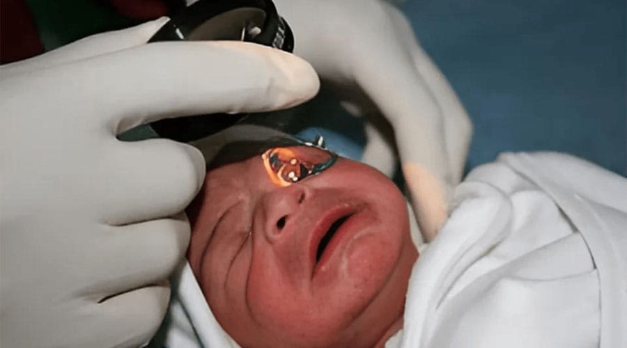
The retina offers a unique window into the body’s blood vessels. In premature infants, doctors routinely capture retinal images to screen for retinopathy of prematurity, a condition that can cause blindness.
Using these existing images, researchers trained an artificial intelligence model to detect subtle vascular patterns associated with lung and heart disease. According to the study published by the University of Colorado Anschutz Medical Campus, the algorithm identified patterns that correlate with serious cardiopulmonary complications.
This approach builds on a growing body of evidence that retinal imaging can reveal systemic disease. Previous research has shown AI systems can detect cardiovascular risk factors from eye images, including age, smoking status, and blood pressure.
In this case, the AI analysis of eye photos leverages data already collected in neonatal intensive care units. That means no additional invasive tests for fragile infants.
Why Early Detection Matters for Premature Infants
Each year, an estimated 15 million babies are born prematurely worldwide, according to the World Health Organization. Many of them require intensive respiratory support.
Conditions like bronchopulmonary dysplasia and pulmonary hypertension can progress quickly. Traditional diagnosis often relies on clinical signs, imaging, and specialist evaluations that may occur after damage has begun.
If AI analysis of eye photos can flag high risk infants earlier, clinicians could intervene sooner. Earlier treatment may improve outcomes and reduce long term complications, including chronic lung disease.
Benefits and Ethical Considerations
The potential advantages are clear:
- Non invasive screening
- Faster risk assessment
- Better allocation of NICU resources
However, AI in neonatal care raises important questions. Algorithms must be rigorously validated across diverse populations. Bias in training data could lead to unequal performance. Clinical decisions should not rely solely on automated predictions.
Experts consistently emphasize that AI should support clinicians, not replace them. Transparency in how models are trained and tested is essential to maintain trust.
What This Means for the Future of Neonatal AI
The broader implication is significant. The AI analysis of eye photos represents a shift toward multi purpose medical imaging, where one diagnostic test serves multiple functions.
For hospitals, this could mean improved efficiency. For families, it could mean earlier answers during an emotionally intense time.
While further validation and peer reviewed studies are needed, this research points to a future where artificial intelligence augments frontline neonatal care with precision and speed.
Conclusion
The idea that a simple retinal image could help detect severe lung and heart disease in premature infants once sounded improbable. Today, it is increasingly plausible.
As artificial intelligence continues to mature, its integration into neonatal medicine may help clinicians act faster, reduce risk, and ultimately save lives. The key will be careful validation, ethical deployment, and keeping human judgment at the center of care.
Fast Facts: AI Eye Diagnosis Explained
What is the new AI method for detecting lung and heart conditions in premature infants?
Researchers developed an artificial intelligence model that analyzes routine retinal images taken during eye exams to detect patterns associated with serious lung and heart diseases in premature babies. This approach could identify conditions earlier without extra procedures.
What can AI analysis of eye photos detect?
The AI system showed it can predict bronchopulmonary dysplasia (a chronic lung disease) and pulmonary hypertension (a dangerous form of high blood pressure affecting the lungs and heart) with high accuracy using retinal images taken during retinopathy screening.
Why is this development significant for neonatal care?
AI analysis of eye photos is significant because eye imaging is already part of standard retinopathy of prematurity (ROP) screening, using AI to analyze those images could enable earlier detection of life-threatening complications and reduce reliance on invasive diagnostic testing..